Preparate microscopice
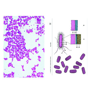
Componentele de bază ale sistemului sunt cele patru serii de preparate microscopice pentru școli A, B, C și D. Seriile sunt ordonate sistematic și alcătuite astfel încât fiecare serie se bazează pe cea precedentă și extinde domeniul de cunoștințe al seriei anterioare. Fiecare preparat este selectat cu grijă și verificat din punct de vedere al valorii sale didactice. La selectarea preparatelor s-a acordat prioritate celor tipice pentru grupul de animale sau plante corespunzător. Materialul este disponibil în cantități suficiente, astfel încât profesorul să poată selecta și varia. Preparatele micro LIEDER sunt fabricate în laboratoarele noastre sub conducere științifică. Ele sunt rezultatul unei experiențe de zeci de ani în toate domeniile tehnicii de preparare. Secționarea microtomică este efectuată de specialiști cu experiență, tehnica de secționare și grosimea secțiunii sunt adaptate obiectelor. Din numărul mare de metode de colorare utilizate în microscoape, alegem cele care combină o reprezentare clară și contrastantă a structurilor dorite cu o durabilitate optimă. În majoritatea cazurilor, este vorba de colorări multiple complexe. Preparatele microscopice LIEDER sunt livrate pe lamele fin șlefuite în format 26 x 76 mm. Fiecare preparat microscopic este unic. Prin urmare, dorim să atragem atenția asupra faptului că preparatele livrate pot diferi de ilustrațiile din acest catalog, din cauza variațiilor naturale ale materialelor de bază și a metodelor de preparare și colorare utilizate.
Numărul seriei de preparate disponibile, sau cel puțin al unor părți din acestea, trebuie să corespundă aproximativ cu numărul de microscoape disponibile, astfel încât mai mulți elevi să poată examina simultan aceleași preparate. Prin urmare, toate preparatele microscopice din serii pot fi achiziționate și individual, astfel încât preparatele importante să poată fi achiziționate în cantități suficiente pentru întreaga clasă.
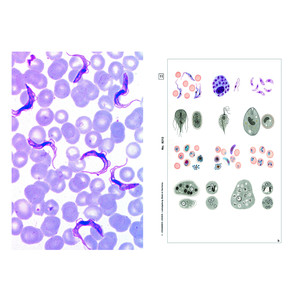
Parazitologie, 50 preparate
Paraziți indigeni și tropicali ai omului și animalelor domestice
1. Entamoeba histolytica, diaree amoebică
2. Leishmania donovani, agent patogen al Kala-Azar
3. Trypanosoma gambiense, boala somnului, frotiu de sânge
4. Trypanosoma cruzi (Schizotrypanum), boala Chagas
5. Plasmodium falciparum, malarie tropicală la om, frotiu sanguin cu stadii inelare
6. Plasmodium berghei, malarie la rozătoare, frotiu sanguin
7. Plasmodium, splina umană cu malarie melanoidă, transversal
8. Toxoplasma gondii, toxoplasmoză
9. Babesia canis, agent patogen al piroplasmozei
10. Sarcocystis sp., în țesutul muscular. Tuburi Miescher
11. Nosema apis, loacă de albine, intestin de albină transversal
12. Monocystis agilis, gregarine din viermele de pământ
13. Eimeria stiedae, coccidioza iepurilor, secțiune prin ficat
14. Fasciola hepatica, fluke mare, total
15. Fasciola hepatica, centrul corpului, transversal
16. Fasciola hepatica, ouă
17. Fasciola hepatica, miracidii (larve ciliate)
18. Schistosoma mansoni, bilharziosis, mascul sau femelă, total
19. Schistosoma mansoni, redii și cercarii în ficatul infectat al melcilor
20. Schistosoma mansoni, ouă în fecale
21. Taenia sau Moniezia, tenie, scolex (cap) cu ventuze, total
22. Taenia pisiformis, vierme plat al câinilor, proglottide mature (segmente), total
23. Taenia saginata, vierme plat al bovinelor, proglottide, transversal
24. Taenia saginata, ouă
25. Hymenolepis sp., vierme plat pitic, proglottide total
26. Echinococcus granulosus, vierme plat al câinilor, scolices (capete) cu coroană de cârlige, total
27. Echinococcus granulosus, perete chist (hidatidă), transversal
28. Ascaris lumbricoides, vierme rotund, regiunea genitală a femelei, transversal
29. Ascaris lumbricoides, regiunea genitală a masculului, transversal
30. Ascaris lumbricoides, ouă
31. Enterobius vermicularis (Oxyuris), vierme intestin, total
32. Trichinella spiralis, trichinelă, larve în mușchi, secțiune transversală
33. Ancylostoma, vierme cu cârlig, mascul sau femelă, total
34. Trichuris trichiura, vierme bici, ouă
35. Strongyloides, vierme filiform pitic, larve totale
36. Heterakis spumosa, parazit la rozătoare, total
37. Ixodes, căpușă, imago, total. Vectori ai encefalitei și borreliozei
38. Dermanyssus gallinae, acarian de găină, total
39. Acarapis woodi, Varroa, acarianul albinelor, total
40. Sarcoptes, acarianul pruritic, secțiune de piele infestată
41. Stomoxys, țânțar de picioare, aparate bucale înțepătoare-sugătoare
42. Anopheles, țânțar malaric, cap și aparate bucale ale femelei
43. Culex pipiens, țânțar, cap și părți ale gurii femelei
44. Anopheles, țânțar purtător de malarie, larvă
45. Culex pipiens, țânțar, larvă
46. Culex pipiens, pupă
47. Cimex lectularius, păianjen de pat,
48. Pediculus humanus, păduche de cap sau de corp
49. Pediculus humanus, ouă de păduchi de cap pe păr (lindini)
50. Ctenocephalus canis, purice de câine
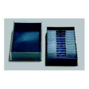
În livrare și în preț sunt incluse unul sau două recipiente din plastic pentru câte 25 de preparate. Se pot comanda suplimentar alte cutii de depozitare.
